Part of the Microscopy series:
- Early History of Microscopy
- Lenses and Image Formation
- Objective lenses & Aberrations
- Diffraction & Interference
- Point Spread FunctionThis post!
- Resolution & Sampling
- Fourier Optics
Point Spread Function
Effect of All the Wavelets: the PSF
Finally, let’s put them all in:
 Effect of all the wavelets - PSF1
This is basically an infinite number of wavelets, and this distribution is now the point spread function.
What we get in the middle is a very elliptical shape, (which should have been a nice symmetrical sphere), and a lot of light is still left around the area of the ideal object. This is actually the limitation of imaging with a light microscope.
Effect of the NA/wavelength on Fringes
- high NA: small central peak, narrow fringes
- low NA: large central peak, wide fringes

- short wavelength: small PSF

PSF Light Distribution Near the Image Plane (XY and XZ)
Looking down on the in-focus plane (XY)
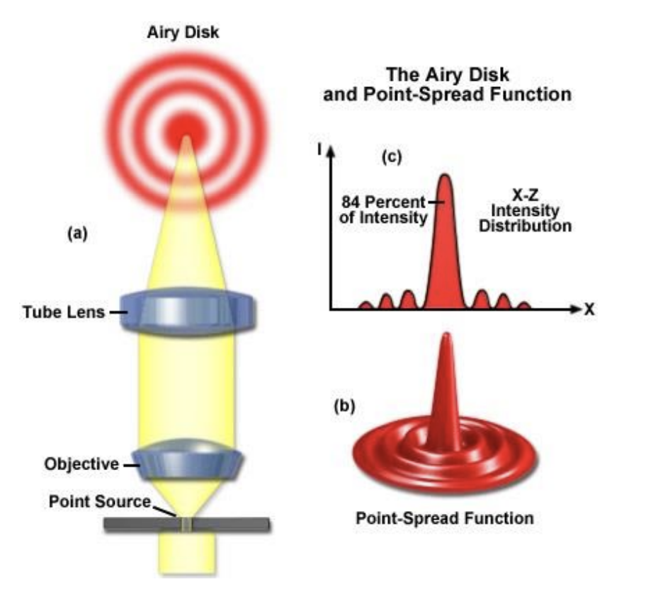
- PSF has center Airy disk;
- PSF has a series of concentric rings.
- Larger rings have progressively lower intensity;
- The first dark ring radius is $0.61\frac{\lambda}{NA}$.
 The Airy Disk and PSF2

On an XZ section
- More spread along the optical axis (Z axis);
- First dark island at $2n\frac{\lambda}{NA^2}$; ( $n$ is the refractive index.)
- Most light within two cones.

Overall distribution

Width and Depth of PSF3
Effect of NA on PSF
Bigger effect on axial (X-Z) than lateral (X-Y) spread.

Convolution
The microscope optics convolve each point source in the specimen with the PSF to produce the image.
$$ Specimen \otimes PSF = Image $$

Objects in a diffraction-limited image of your sample will never appear smaller than the PSF.
For example, that’s say you have two microtubules in your specimen and you have labeled them with GFP, and you imaged them with a 1.4 NA oil objective lens. Microtubules are about 25 nm in diameter, and the PSF for this setup is about 240 nm. So each fluorophore in the microtubule will be convolved with the PSF. You can easily tell by the end of the microtubule that there are two separate microtubules, but not at all by the middle where they are closer than the PSF4.
